
In Class
‘Difficult to Diagnose’
Medical students and residents learn some of the educational power of an autopsy. By Scott Hauser
 |
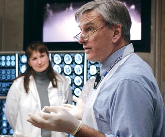
| CONFERENCING: Resident Zsdral (left) and Powers during a recent session (Photo by Elizabeth Torgerson-Lamark) |
Jim Powers is well versed in the anatomical details of the brain he’s about to cut open. The mass of corrugations and ridges that to nonmedical eyes seem indistinguishable are familiar pieces of a biological puzzle that has intrigued the professor of pathology for more than 30 years.
But before he begins the dissection, Powers wants to know some of the details about the person whose death was caused by a disease that has left telltale clues somewhere below the folds of the brain that now lies on the table.
In this case, that was a young man who was diagnosed with a brain tumor when he was 12 years old and who, after nearly seven years of treatment, died last winter at age 18.
“This is a case of a poor little boy,” Powers begins, as he goes over the clinical details in front of a semicircle of medical students and residents. The young man’s doctors believe the cause of death was gliomatosis cerebri—a particularly “bad tumor,” Powers notes—that’s known for infiltrating throughout the tissue of the brain.
“It’s an easy lesion to see on autopsy but difficult to diagnose in a surgical biopsy,” Power says.
And that’s why Powers and the students have gathered in a pathology lab in the School of Medicine and Dentistry, as they do nearly every week during the academic year. Formally called the Brain Cutting Conference, the session is part of the neurology rotation, in which students spend 10 weeks learning about the brain and its functions.
The conference, a four-week part of the rotation, gives students and residents a chance to see firsthand the damage that disease wreaks on the brain.
For Powers, who also is a professor of neurology, the sessions are important teaching moments that can link clinical care—seeing patients at the bedside and treating their ailments—and pathology—understanding the mechanisms that damage human tissue.
“The conference really helps to consolidate things,” Powers says later. “For medical students, it helps to reinforce what they’ve been taught in their courses. For pathology residents, it helps reinforce what they’ve already been exposed to, and it introduces them to some new entities.
“And for the clinicians who sometimes attend, it can help confirm or disprove a diagnosis,” he says. “It really helps put things together.”
Krisztina Zsdral, a pathology resident, says the perspectives brought to the conference by physicians—who sometimes knew the patient—as well as radiologists, pathologists, and students make the session especially interesting.
“The clinicians talk about what they see, and the radiologists talk about what they see,” Zsdral says. “It puts together the whole picture.”
And as he leads the session, Powers, who specializes in the pathology of the brain and directs the neuropathology and postmortem medicine services at Strong Memorial Hospital, offers a glimpse of how pathologists approach their work.
He’s careful to point out that definite answers are often elusive, noting that pathologists usually provide a probable cause of death rather than a definitive one.
“We’re consultants,” he says to the students and residents. “We’re diagnostic specialists, but you need to know our limits.”
Allen Pardee, a neurology chief resident who oversees the medical students during their rotation, says the conference provides an important perspective for students in an age when medical imaging techniques have made diagnosing brain damage more exact.
“The pictures don’t always convey the dramatic impact in the same way as seeing it in the conference,” Pardee says.
The conference, Powers says, also is a chance to introduce students to the diagnostic power provided by autopsies.
“I’m a big believer in autopsies,” he says. “It’s probably the most underappreciated resource we have.”
Autopsies are becoming increasingly less appreciated, with fewer full autopsies requested by both doctors and families each year, Powers says.
The trend is driven by increasing reliance on imaging technology, changes in the way pathology services are calculated by Medicare, and a lack of education on the part of the public, but also by declining interest among medical professionals, who often think autopsies are unnecessary to understand a cause of death.
Even among pathologists, interest is waning, Powers says, due to several factors, not least of which is the amount of work involved. A full autopsy often takes three or four days of arduous work—“It’s quite literally a visceral experience,” Power says—and completing an autopsy report can take weeks.
But he says the value of examining diseased tissue is vital to understanding not only causes of death, but also in providing new information to help understand disease mechanisms. And for some families, autopsies can offer a sense of closure and a sense that their loved one’s death may lead to clues to preventing future deaths.
“Sometimes family members will say, ‘You guys didn’t know anything about this disease, and we’d like some good to come out of this,’” he says. “The family can have some consolation, if you want to call it that, of being able to contribute some help to the next family that has to face a particular disease.”
