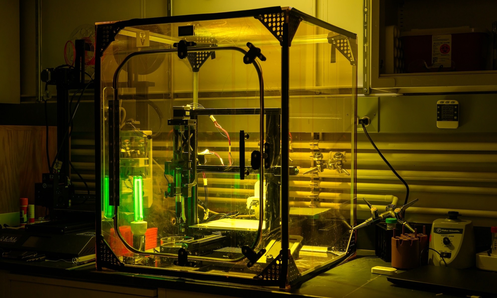
URochester researchers are developing noninvasive, light-based imaging to spot fractures that aren’t healing properly, without relying on frequent X-rays.
About 6.8 million Americans get treated for broken bones every year, and while most fractures heal on their own, about 10 percent go awry, which can lead to risks of refracture, extended and painful recoveries, or even permanent disability. Doctors use X-rays to monitor bone healing but must limit how frequently they use the imaging technique so that patients are not exposed to unhealthy amounts of radiation.
Researchers at the University of Rochester are developing non-invasive optical techniques that do not expose patients to the same kind of radiation that makes frequent X-rays a concern. These techniques would help detect early on whether a bone is healing properly, and if not, predict the right treatments to accelerate the healing process. Regine Choe, the Marylou Ingram Professor in Biomedical Engineering, and members of her lab have worked on a method using near-infrared light since 2018.
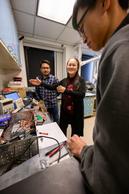
Choe recently received nearly $3 million from the National Institutes of Health to advance the technology toward clinical studies involving people with a notoriously problematic injury: the Jones fracture.
Shedding light on a gray area
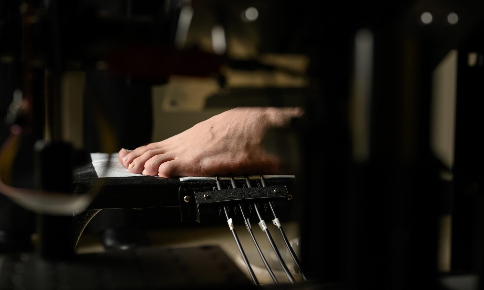
Jones fractures occur when a person breaks the fifth metatarsal—the long bone on the outside of the foot connected to the pinkie toe. Restoring blood flow to fractured bones is critical for proper healing, and Choe says the fifth metatarsal is a common place where complications arise.
“The bone is right in a sweet spot where the blood vessels often don’t provide enough blood supply to properly heal the foot,” says Choe. “But it’s hard to predict—sometimes it heals well and sometimes it doesn’t. We want to use non-invasive imaging to clear up that gray area.”
Choe’s lab uses closely related techniques called diffuse optical tomography and diffuse correlation tomography. Essentially, the researchers use near-infrared light, which falls in the region of the electromagnetic spectrum just beyond visible red light. Directed at the injured foot in a series of small beams that cover a few inches, the light penetrates beneath the skin, allowing researchers to collect information about blood volume, the blood’s oxygen content, and blood flow rate.
“That information can tell us whether blood flow is restored after the bone fracture,” says Choe. “And since we’re using light, it is safe to make frequent measurements to see how well revascularization is progressing.”
Taking the next step
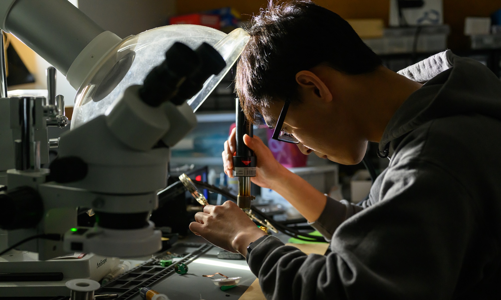
Choe and her students have developed a prototype device that they intend to refine and begin testing on patients with Jones fractures with support from clinical collaborators at University of Rochester Medicine.
The researchers envision their technique as more than just a monitoring tool. They intend to make predictions about how well a bone will heal and account for differences in patients such as age and gender. When a bone is not healing correctly, they also want to use their technique to optimize the right dosage and timing for treatments such as parathyroid treatment, which is already used to treat osteoporosis but is not yet used for fractures.
“Some researchers have tried using parathyroid hormone for fracture healing, but there is some discrepancy between dosage, timing, and other factors that need to be resolved for it to be applicable,” says Choe. “We think our method can help optimize treatments being tested for fractures that do not heal well.”
The study is expected to be completed in 2031.